TEE Colour Doppler app for iPhone and iPad
Developer: University Health Network
First release : 24 Nov 2013
App size: 64.46 Mb
Two and three-dimensional echocardiography forms the basis of assessing cardiac structure and function. The application of colour Doppler imaging is routinely performed over cardiac valves, vascular and other pathological structures to assess blood flow direction and velocity. This aids the confirmation of pathological diagnoses and quantifies the severity of these lesions.
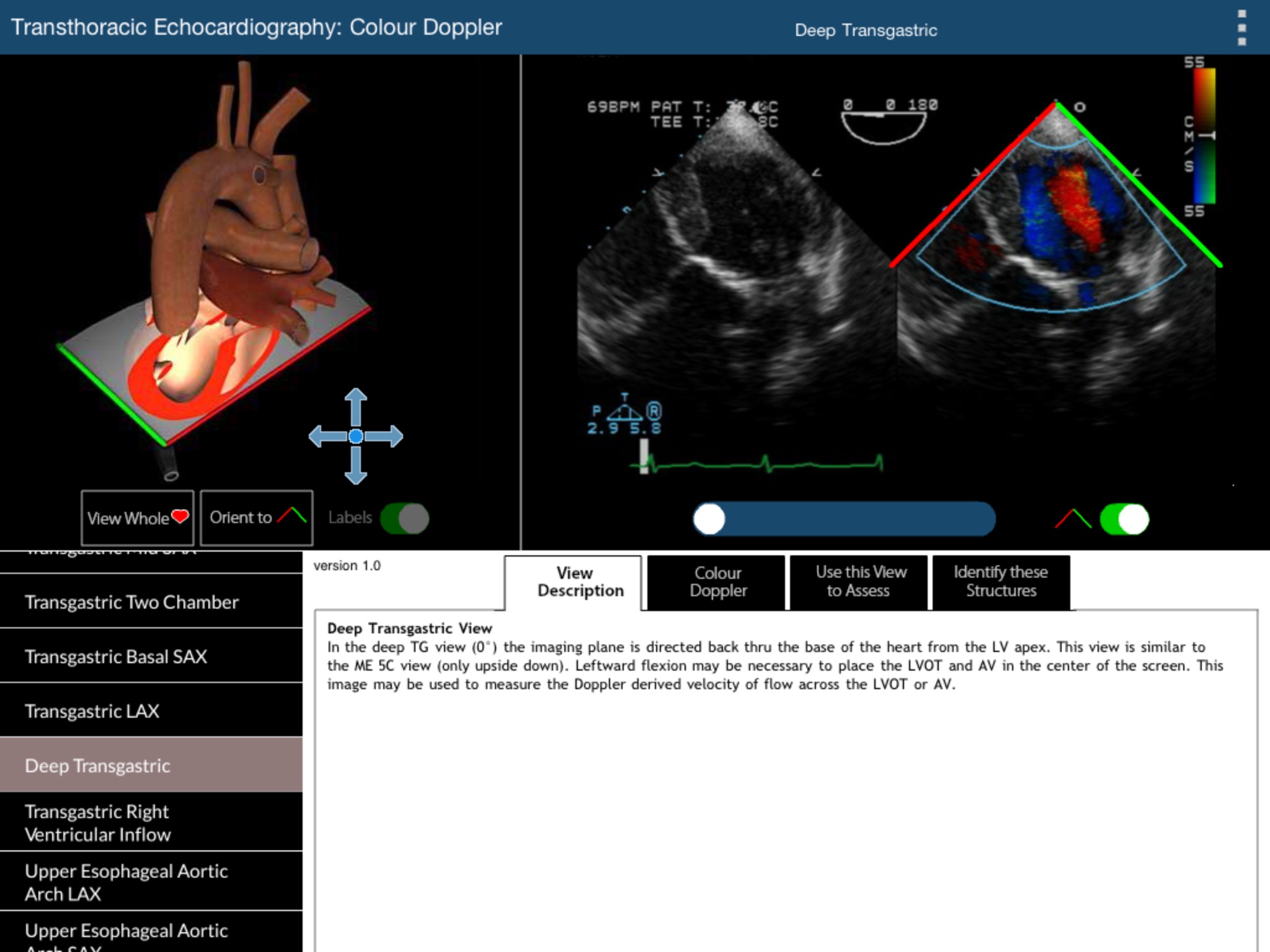
This module demonstrates which structures should be viewed using colour Doppler during the standard 2D examination. Each of the 20 standard views is accompanied by a rotatable three-dimensional heart model and a `colour compare’ video clip which simultaneously shows the two-dimensional and colour Doppler images of each view. Normal blood flow across or within various parts of the heart is shown.


